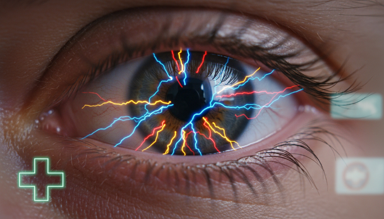
Photopsia, the medical term for flashing lights in the eyes, describes brief visual disturbances that appear as sparks, streaks, or lightning bolts across your field of vision. These sensations occur when the retina—the light-sensitive tissue at the back of the eye—experiences mechanical stimulation from shifting vitreous gel or neurological events in the visual cortex.
While often harmless, particularly in older adults experiencing natural aging changes, these flashes sometimes signal serious conditions requiring urgent intervention. Understanding the distinct patterns—from peripheral arcs to zigzag lines—helps distinguish between benign posterior vitreous detachment and sight-threatening retinal tears or vascular emergencies.
Why am I seeing flashes of light in the corner of my eye?
- Flashes in the corner or periphery most commonly indicate PVD, where the vitreous gel separates from the retina.
- Repeated bursts of light in one eye suggest possible retinal traction or tearing.
- The risk of retinal detachment increases when flashes accompany a sudden shower of floaters.
- Older adults face higher incidence due to age-related vitreous shrinkage.
- Pattern distinction matters: unilateral sparks differ significantly from bilateral zigzag patterns.
- While many cases resolve without intervention, some require emergency surgical repair.
| Symptom Pattern | Likely Cause | Urgency Level |
|---|---|---|
| Corner or peripheral flashes | Posterior vitreous detachment | Monitor; seek care if new |
| Zigzag lines both eyes | Migraine with aura | Low if typical history |
| Sudden flashes in one eye | Retinal tear or detachment | Emergency |
| Arc-shaped flashes | Vitreous traction on retina | Schedule dilated exam |
| Flashes with curtain/shadow | Retinal detachment | Immediate intervention |
| Brief sparks with floaters | Vitreous gel changes | Routine evaluation |
What causes zigzag flashing lights in the eyes?
Zigzag patterns, shimmering arcs, and geometric shapes crossing the visual field typically originate from the brain rather than the retina. According to Dark and Refocus Eye Care, these visual phenomena represent migraine aura—a neurological event affecting both eyes simultaneously, usually lasting between five and sixty minutes.
Unlike retinal flashes, which appear as brief sparks or streaks in one eye’s periphery, migraine auras present as bilateral distortions that may precede or accompany headache pain. Bright Sight Eye Clinic notes that these patterns differ fundamentally from mechanical retinal stimulation by their duration and symmetric presentation across both visual fields.
How do migraine auras differ from retinal flashes?
Retinal flashes result from physical tugging on photoreceptor cells, producing momentary lightning-like sensations typically limited to one eye. Migraine auras, conversely, represent cortical spreading depression—a wave of electrical activity moving across the visual cortex. This creates the characteristic fortification spectra or zigzag patterns visible in both eyes.
Healthline emphasizes that migraine-related flashing lights rarely accompany the floating specks or cobwebs associated with vitreous detachment, providing a key diagnostic differentiator for clinicians assessing sudden visual changes.
Migraine auras typically resolve within an hour and affect both eyes simultaneously. Retinal flashes persist for milliseconds, recur unpredictably, and usually localize to one eye’s peripheral vision. New onset zigzag patterns without previous migraine history warrant retinal examination to exclude alternative pathology.
What does it mean if flashing lights appear in one eye?
Unilateral flashing lights—confined to a single eye—most frequently indicate mechanical events within the vitreous chamber. Duke Health explains that as the vitreous gel ages, it liquefies and shrinks, eventually pulling away from the retinal surface in a process called posterior vitreous detachment.
This separation creates friction against the retina, generating the perception of sparks or lightning streaks in the affected eye’s periphery. While PVD occurs commonly in individuals over 50 and usually resolves without complication, the traction can occasionally tear the retinal tissue, creating pathways for fluid accumulation beneath the retina.
When does unilateral flashing indicate emergency?
AARP Health warns that repeated bursts of light combined with a sudden increase in floaters, peripheral shadows, or curtain-like vision loss signal retinal detachment—a medical emergency risking permanent vision loss. The Northwest Eye Clinic confirms that immediate surgical intervention offers the best prognosis for preserving sight when detachment occurs.
Trauma represents another cause of unilateral symptoms. Physical injury can cause immediate vitreous traction or retinal damage, producing flashes and floaters that require urgent ophthalmologic assessment to assess structural integrity.
Why do I see flashing lights in my eyes when closed or trying to sleep?
Visual disturbances observed in darkness or during sleep onset often reflect the same underlying mechanisms active during waking hours, though heightened awareness in low-light conditions may increase perception. NHS guidance indicates that PVD-related flashes frequently persist when the eyes are closed, as the mechanical stimulation of retinal cells occurs independently of external light input.
During pregnancy, hormonal fluctuations may accelerate vitreous changes, potentially increasing the incidence of photopsia. While direct research on pregnancy-specific flashing lights remains limited, the mechanical principles of vitreous gel alteration appear consistent with physiological changes affecting connective tissue throughout the body.
Do closed-eye flashes require different evaluation?
The character of the flashes matters more than the lighting conditions under which they appear. Brief, arc-like sensations in the periphery suggest vitreous traction, while prolonged geometric patterns point toward neurological origins. Cleveland Clinic specialists recommend that any new onset of flashes, regardless of when they occur, warrants professional evaluation to rule out retinal pathology.
Seek immediate medical attention if flashing lights accompany a sudden increase in floaters, peripheral shadows, or blurred vision. These symptoms may indicate retinal detachment requiring emergency surgical intervention to prevent permanent vision loss.
While pregnancy may heighten awareness of existing vitreous changes due to hormonal effects on connective tissue, specific data on pregnancy-induced photopsia remains limited. Monitor for increases in frequency and consult specialists if patterns change suddenly.
How do flashing lights in eyes progress over time?
The temporal evolution of photopsia follows predictable patterns based on underlying etiology, particularly regarding age-related vitreous changes.
- Vitreous shrinkage initiation: Beginning around age 50, the gel-like vitreous humor gradually liquefies and contracts, creating increasing tension on retinal attachment points.
- PVD onset: As separation from the retina begins, patients typically experience weeks of intermittent flashes in the peripheral vision, often described as sparks or streaks.
- Potential complication: If traction exceeds retinal adhesion strength, a tear may develop, producing a sudden shower of new floaters alongside increased flash frequency.
- Resolution or escalation: Most PVD cases stabilize within months as the vitreous completes its separation, though some patients continue experiencing occasional flashes indefinitely without complication.
Which flashing light symptoms are harmless versus emergencies?
Distinguishing between benign age-related changes and sight-threatening conditions requires careful attention to associated features and symptom evolution.
Established as Generally Benign
- Isolated posterior vitreous detachment in adults over 50
- Brief, occasional sparks without vision changes
- Longstanding stable patterns with no new floaters
- Bilateral zigzag patterns consistent with migraine history
Requires Further Investigation
- Specific correlations between pregnancy stage and flash intensity
- Whether sleep position affects vitreous traction patterns
- Precise thresholds distinguishing normal from excessive flash frequency
- Individual variation in retinal adhesion vulnerability
What physiological mechanisms create flashing lights in eyes?
The perception of light without external photonic input stems from two primary mechanisms: mechanical stimulation of retinal photoreceptors and aberrant neurological signaling in the visual pathway. In posterior vitreous detachment, the collagenous framework of the vitreous humor shrinks and pulls away from the retinal surface, creating friction that depolarizes rod and cone cells. The brain interprets this electrical discharge as light, typically localized to the peripheral retina where separation first occurs.
Migraine auras operate through entirely different pathways. Cortical spreading depression—a slowly propagating wave of neuronal suppression across the visual cortex—creates the characteristic scintillating scotomas and zigzag patterns. Unlike retinal phenomena, these manifestations affect both eyes because they originate in the brain’s processing centers rather than unilateral peripheral sensors.
The association with systemic conditions such as Blood Clot in Lung or other vascular events remains an important consideration in differential diagnosis, as photopsia can occasionally signal vertebrobasilar insufficiency or transient ischemic attacks affecting the visual pathways.
What do medical authorities say about eye flashes?
“Floaters and flashes are usually harmless. If you have them, you should see an optician to check if you have posterior vitreous detachment (PVD) or something more serious like a retinal detachment.”
— NHS Symptom Guidance
“Flashes of light are pinpricks or spots of light that you see in your field of vision. Light flashes can also look like jagged lines or appear wavy. If the vitreous gel shrinks or pulls away from the retina, you may see flashes of light.”
— Cleveland Clinic Medical Review
What should you remember about flashing lights in your eyes?
Flashing lights in the eyes typically originate from benign age-related vitreous changes, but the specific pattern—unilateral sparks versus bilateral zigzags—guides urgency. New onset symptoms, particularly when accompanied by floaters or vision shadows, require immediate specialist evaluation to exclude retinal detachment. While migraine auras resolve spontaneously and PVD stabilizes over months, understanding these distinctions ensures appropriate response to visual changes. For those experiencing systemic symptoms alongside visual disturbances, consider evaluation for conditions like Symptoms of Stomach Cancer or other neurological concerns.
Frequently Asked Questions
What is photopsia?
Photopsia refers to the perception of flashes, sparks, or arcs of light without external light sources, resulting from mechanical retinal stimulation or neurological events.
Are flashing lights always serious?
No. Many cases, particularly in older adults, represent harmless posterior vitreous detachment. However, sudden onset with floaters or vision loss requires emergency evaluation.
How common are eye flashes?
Extremely common in adults over 50, with most people developing some degree of vitreous detachment and associated photopsia as they age.
Can stress cause flashing lights?
Stress may trigger migraine auras with zigzag patterns, but mechanical flashes from vitreous traction are not stress-induced.
Do flashing lights mean I’m having a stroke?
Rarely. While vertebrobasilar insufficiency can cause bilateral flashes, most photopsia stems from ocular rather than cerebral vascular events.
How long do PVD flashes last?
Flashes from posterior vitreous detachment typically persist for weeks to months during the separation process, then diminish as the vitreous fully detaches.
Can dehydration cause eye flashes?
No direct evidence links dehydration to photopsia, though severe dehydration may exacerbate migraine susceptibility.

Oliver Jack Carter Cooper is a staff writer for EveningLedger.uk, covering UK news, politics, business and culture. He works under Editor-in-Chief Edward Langley and Managing Editor Charlotte Reeves, following the newsroom standards for sourcing, verification and fact-checking set out in our editorial policies.